Bai Shao: The Art of Softening the Liver
From the peony roots of East Asia to the cellular processes involved in liver fibrosis.
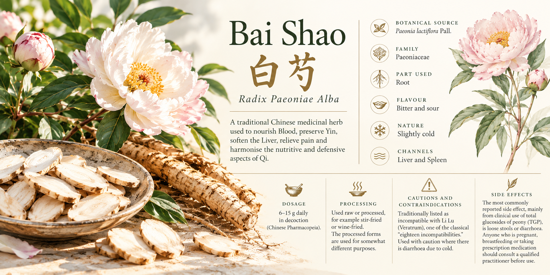
Bai Shao (白芍), the root of Paeonia lactiflora, has a long history of use in Chinese medicine, where it is traditionally described as nourishing Blood, preserving Yin, relieving pain and softening the Liver. It is found in formulas used for menstrual disorders, muscular cramps, abdominal pain and conditions associated with tension or insufficient nourishment of the sinews.
One of its principal chemical constituents, paeoniflorin, has also attracted attention in modern pharmacological research. Laboratory and animal studies suggest that it can influence several processes involved in liver inflammation and fibrosis, including the activation of hepatic stellate cells, which are responsible for much of the excess collagen deposited during liver scarring.
The relationship between these findings and the traditional description of softening the Liver is an interesting subject for enquiry. The two expressions arise from different medical frameworks, and refer to different observations, but both provide an opportunity to examine how the liver responds to changing conditions.
The plant and its natural home
Paeonia lactiflora Pall. is a herbaceous perennial belonging to the Paeoniaceae family. Its natural range extends from southeastern Siberia through Mongolia and into northern, central and eastern China, where it grows in temperate environments.
Unlike the woody tree peonies, its above-ground stems die back each year. During the colder months, the plant survives through its underground structures, producing new shoots when the growing season returns.
Its leaves are divided into smaller leaflets, and the flowers of wild plants are generally relatively simple, often white or pale pink, with a prominent cluster of yellow stamens. The large, densely petalled peonies commonly seen in gardens are the result of generations of horticultural selection.
The root is thick and fleshy, storing reserves accumulated over successive growing seasons. It is this underground part of the plant that provides the medicinal material known as Bai Shao.
The species is recorded by the Royal Botanic Gardens, Kew, as native to regions including Mongolia, Inner Mongolia, Manchuria, parts of the Russian Far East and several areas of China. It is now cultivated more widely for both ornamental and medicinal purposes.
A plant with several names
Peony root has a documented place in Chinese, Korean and Japanese medicine.
In China, the medicinal root is known as Shao Yao (芍藥), with Bai Shao (白芍) and Chi Shao (赤芍) distinguished as different medicinal materials.
In Korea, peony root is known as jakyak (작약, 芍藥). It appears in traditional Korean medicine, including formulas used for abdominal pain, menstrual complaints and muscular spasms.
In Japan, the root is called shakuyaku (芍薬). It is an established Kampo ingredient and appears in several frequently prescribed formulas, including Shakuyakukanzoto, used for painful muscle cramps.
These traditions share historical connections, although their clinical systems and approaches to prescribing have developed in different ways.
The geographical range of the plant also includes Mongolia and parts of southern Siberia, where numerous communities have longstanding traditions of medicinal plant use. However, the presence of a species within a region does not establish its particular use by the people living there. Documented local uses of Paeonia lactiflora would require more specific ethnobotanical evidence.
Bai Shao: a herbal monograph
Bai Shao 白芍, Radix Paeoniae Alba
Botanical source: Paeonia lactiflora Pall.
Family: Paeoniaceae
Part used: Root
Flavour: Bitter and sour
Nature: Slightly cold
Channels: Liver and Spleen
Dosage and cautions
Dosage: 6–15 g daily in decoction (Chinese Pharmacopoeia).
Processing: Used raw or processed, for example stir-fried or wine-fried. The processed forms are used for somewhat different purposes.
Cautions and contraindications: Traditionally listed as incompatible with Li Lu (Veratrum), one of the classical "eighteen incompatibilities." Used with caution where there is diarrhoea due to cold.
Side effects: The most commonly reported side effect, mainly from clinical use of total glucosides of peony (TGP), is loose stools or diarrhoea. Anyone who is pregnant, breastfeeding or taking prescription medication should consult a qualified practitioner before use.
Traditional actions
Nourishes Blood, preserves Yin, softens the Liver, relieves pain and harmonises the nutritive and defensive aspects of Qi.
In Chinese herbal medicine, Bai Shao is particularly associated with the nourishment of Liver Blood and the easing of tension in the sinews.
The expression Yang Xue Rou Gan (養血柔肝), meaning to nourish Blood and soften the Liver, describes one of its principal traditional actions.
Within Chinese medicine, the Liver encompasses a broader range of physiological relationships than the anatomical organ alone. It is associated with the storage of Blood, the smooth movement of Qi, the nourishment of the sinews and aspects of menstrual regulation.
Where Liver Blood is considered insufficient, the sinews may be inadequately nourished, contributing to cramps, spasms or muscular tightness. Bai Shao is traditionally used to address these patterns, often in combination with other herbs.
One familiar example is Shao Yao Gan Cao Tang, a two-herb formula combining Bai Shao with Gan Cao, liquorice root. It is traditionally used for cramping and spasmodic pain.
Bai Shao also appears in Si Wu Tang, alongside Shu Di Huang, Dang Gui and Chuan Xiong, in a classical formula used to nourish and regulate Blood.
In Xiao Yao San, it is combined with herbs including Chai Hu, Dang Gui and Bai Zhu in a formula traditionally used for patterns involving constrained Liver Qi and Blood deficiency.
The therapeutic role of Bai Shao varies according to the formula, its preparation and the other herbs with which it is combined.
Bai Shao and Chi Shao
White peony root, Bai Shao, is distinguished from red peony root, Chi Shao (赤芍).
Both can be derived from Paeonia lactiflora, although Chi Shao can also be obtained from Paeonia veitchii.
Their preparation differs. Bai Shao is commonly produced from cultivated roots that are cleaned, boiled, peeled and dried, while Chi Shao is generally dried without the same boiling and peeling process.
The two are also understood differently in Chinese medicine.
Bai Shao is associated with nourishing Blood, preserving Yin and relieving tension, while Chi Shao is more strongly associated with cooling Blood and dispersing Blood stasis.
Although they share some chemical constituents, they are recognised as distinct medicinal materials.
The chemistry of the root
Chemical analysis of Paeonia lactiflora has identified numerous compounds, including monoterpene glycosides, phenolic compounds, flavonoids and tannins.
Among the most extensively investigated are paeoniflorin and albiflorin.
Paeoniflorin is a monoterpene glycoside and an important marker in the quality assessment of peony root. It has been studied for its effects on inflammation, oxidative stress, immune signalling and cellular activity.
Researchers also investigate preparations known as total glucosides of peony, or TGP, which contain a mixture of related constituents, including paeoniflorin.
These preparations are chemically different from whole Bai Shao root.
An experiment involving isolated paeoniflorin cannot automatically be taken as evidence that a traditional Bai Shao decoction will produce the same effect. The quantities involved, absorption, metabolism and interactions between compounds may all differ.
Nevertheless, studying individual constituents can help identify some of the biological processes that may contribute to the activity of the medicinal plant.
The cells of the liver
The liver contains several types of cells, each contributing to the maintenance and function of the organ.
The principal functional cells are hepatocytes. They account for most of the liver's mass and carry out much of its metabolic work.
Hepatocytes process nutrients, manufacture proteins, produce bile, store and release glucose, metabolise fats and transform substances for use or removal from the body.
They also have a considerable capacity to regenerate. Following injury, surviving hepatocytes can multiply to replace lost cells, provided the surrounding conditions permit recovery.
However, the liver's function depends on more than hepatocytes alone.
Its tissue contains blood vessels, immune cells, supporting cells and a network of proteins and other substances known as the extracellular matrix.
Among the supporting cells are hepatic stellate cells.
Hepatic stellate cells
Hepatic stellate cells are located in a narrow region called the space of Disse, between the hepatocytes and the specialised blood vessels known as liver sinusoids.
In a healthy liver, most stellate cells are relatively inactive, or quiescent.
They store vitamin A in small lipid droplets and participate in maintaining the extracellular matrix surrounding the liver cells.
The extracellular matrix provides structural support and helps organise the relationships between cells. It is continually maintained and remodelled as part of ordinary tissue function.
When the liver is injured, stellate cells receive signals from damaged hepatocytes, immune cells and other parts of the surrounding tissue.
These signals can cause them to become activated.
The cells change their shape, lose much of their stored vitamin A and begin producing larger quantities of collagen and other extracellular matrix proteins.
They acquire characteristics similar to myofibroblasts, the cells involved in wound healing elsewhere in the body.
This is initially a useful response. When tissue is injured, additional structural material helps stabilise the damaged area while repair takes place.
The difficulty arises when the injury continues.
From repair to fibrosis
Chronic liver injury can occur for several reasons, including persistent viral infection, alcohol-related disease, metabolic dysfunction-associated steatotic liver disease and autoimmune liver disorders.
When injury is repeated or sustained, the inflammatory and repair signals may continue for months or years.
Activated hepatic stellate cells respond by producing increasing quantities of collagen, particularly types I and III.
Over time, the extracellular matrix accumulates, and the physical structure of the liver begins to change.
Healthy liver tissue is relatively flexible. Fibrotic tissue becomes progressively stiffer, and the normal organisation of cells and blood vessels may be disrupted.
This change in stiffness can itself influence cellular behaviour.
Cells respond to the mechanical properties of their surroundings through a process called mechanotransduction. Proteins associated with the cell membrane detect forces and resistance in the surrounding tissue and transmit signals into the cell.
A stellate cell surrounded by stiff, collagen-rich tissue may therefore receive both chemical and mechanical signals that encourage it to remain activated.
The activated cell produces more collagen, increasing the stiffness of the surrounding tissue and potentially reinforcing the conditions that maintain its activity.
This process can contribute to the persistence of fibrosis.
The mechanisms involved have been described extensively in research into liver fibrosis and its regression.
Can liver fibrosis regress?
Fibrosis was once widely regarded as an essentially irreversible process, particularly when it had progressed to cirrhosis.
Research has since demonstrated that some fibrotic changes can regress when the underlying cause of liver injury is removed or effectively treated.
This has been observed, for example, in some patients following successful treatment of chronic hepatitis C.
The degree of recovery varies considerably. Advanced cirrhosis can leave lasting changes in liver architecture and blood flow, even when some scar tissue regresses.
Nevertheless, the liver retains an important capacity for repair.
For fibrosis to regress, two broad processes must occur.
First, the production of new scar-forming material needs to diminish.
Second, some of the accumulated extracellular matrix must be broken down and removed.
Macrophages and enzymes involved in matrix remodelling contribute to this process.
The population of activated hepatic stellate cells must also change. Research has identified several possible outcomes for these cells, including apoptosis, inactivation and senescence.
These are distinct biological processes, each with different consequences for the surrounding tissue.
Three possible fates of an activated stellate cell
Apoptosis: programmed cell death
Apoptosis is a controlled process through which a cell dismantles itself.
It is part of ordinary tissue maintenance and allows the body to remove cells that are damaged, unnecessary or no longer receiving the signals required for survival.
During apoptosis, a cell shrinks, its internal structures are broken down and its contents are packaged into fragments that can be cleared by neighbouring cells and immune cells.
Activated hepatic stellate cells can undergo apoptosis when the conditions supporting their activation begin to disappear.
The process is regulated by several groups of proteins, including the Bcl-2 family and enzymes called caspases.
The Bcl-2 family helps determine whether a cell proceeds towards apoptosis, while caspases carry out much of the controlled dismantling.
When activated stellate cells undergo apoptosis, the number of cells producing excessive collagen is reduced.
This does not immediately remove the collagen already present, but it can reduce continued production.
Inactivation: a return towards quiescence
A second possibility is that an activated stellate cell survives but becomes less active.
This process is described as inactivation or reversion towards a quiescent-like state.
The cell reduces its production of collagen and other proteins associated with fibrosis. Its metabolism and gene expression also change.
One important regulator is PPARγ, a protein involved in lipid metabolism and the maintenance of the quiescent stellate-cell phenotype.
PPARγ activity generally decreases when stellate cells become activated. Experimental studies suggest that restoring its activity can encourage cells towards a less fibrogenic condition.
However, inactivation does not necessarily restore the cell to exactly its original state.
Some inactivated stellate cells retain molecular characteristics associated with their previous activation and may respond more readily if injury occurs again.
The cell can therefore become less active without completely losing the effects of its earlier experience.
Senescence: the cell stops dividing
A third possible outcome is cellular senescence.
Senescence is a state in which a cell remains alive and metabolically active but enters a stable arrest of its normal division cycle.
It can arise in response to DNA damage, oxidative stress, repeated cell division and other forms of cellular disturbance.
In activated hepatic stellate cells, senescence can limit further proliferation and reduce fibrogenic activity.
Proteins including p53, p21 and p16 are involved in regulating aspects of this process.
Senescent cells may also release signals that attract immune cells, which can participate in their removal.
However, senescence is not always beneficial.
Senescent cells can produce inflammatory substances and other signalling molecules, collectively described as the senescence-associated secretory phenotype, or SASP.
Depending on the circumstances, these substances may assist tissue repair or contribute to ongoing inflammation.
The consequences of senescence therefore depend on the type of cell involved, the surrounding conditions and whether the senescent cells are eventually cleared.
Apoptosis, inactivation and senescence are recognised features of the biology of fibrosis and its regression, although their relative contributions vary between experimental models and disease conditions.
Bai Shao and the biochemistry of fibrosis
The relationship between paeoniflorin and liver fibrosis has been investigated through several different experimental approaches.
Much of the research concerns the signals that cause hepatic stellate cells to become activated and remain active.
Three areas have received particular attention: TGF-β signalling, oxidative stress and cellular metabolism.
TGF-β: signalling for collagen production
Transforming growth factor beta, usually abbreviated to TGF-β, is one of the principal signalling molecules involved in fibrosis.
When TGF-β1 binds to receptors on a hepatic stellate cell, it activates a series of proteins called Smads.
These proteins help transmit the signal to the cell's nucleus, influencing the expression of genes involved in collagen production and other fibrotic processes.
In 2018, researchers investigated paeoniflorin in rats with radiation-induced liver fibrosis.
They found that treatment reduced collagen deposition and altered the expression of proteins involved in TGF-β1/Smad signalling.
In particular, the study reported reduced Smad3 and Smad4 expression and increased Smad7 expression. Smad7 helps inhibit aspects of TGF-β signalling.
The researchers also observed improvements in several biochemical markers of liver injury.
These findings suggest that paeoniflorin can influence a major pathway involved in the development of fibrosis.
However, a reduction in collagen production or stellate-cell activation does not establish that an activated cell has fully returned to its original quiescent state.
Oxidative stress and HO-1
A second area of research concerns oxidative stress.
Cells naturally produce reactive oxygen species during metabolism. In controlled quantities, these molecules participate in ordinary cellular signalling.
When produced in excess, they can damage proteins, fats and DNA and contribute to inflammatory responses.
Oxidative stress is an important feature of several forms of chronic liver injury and can contribute to hepatic stellate-cell activation.
One protective enzyme involved in the response to cellular stress is heme oxygenase-1, or HO-1.
In a 2021 study, researchers investigated the effects of paeoniflorin in mice with liver fibrosis induced by carbon tetrachloride, a chemical commonly used in experimental models of liver injury.
Paeoniflorin treatment increased HO-1 expression and reduced several markers of oxidative stress and inflammation.
The researchers also found reduced expression of α-smooth muscle actin, or α-SMA, along with collagen-related proteins and other markers associated with stellate-cell activation.
The treated animals showed less fibrotic change than the untreated experimental group.
This suggests that paeoniflorin may influence fibrosis partly by changing the inflammatory and oxidative environment in which stellate cells operate.
The study appears to have begun paeoniflorin treatment while liver injury was still being induced. It therefore provides evidence of reduced fibrosis development under experimental conditions, rather than proof that longstanding scar tissue can be reversed.
Cellular metabolism, HIF-1α and mitochondria
A third area of investigation concerns cellular metabolism.
When hepatic stellate cells become activated, their metabolic requirements change. They must support proliferation, contraction and the production of substantial quantities of extracellular matrix proteins.
Mitochondria are involved in meeting these requirements. They contribute to energy production, metabolic regulation and cellular signalling.
One protein involved in the response to reduced oxygen availability is hypoxia-inducible factor 1 alpha, or HIF-1α.
HIF-1α helps cells adapt to low-oxygen conditions by altering the expression of genes involved in metabolism and survival.
It has also been implicated in the activation of hepatic stellate cells.
In a 2014 study, Zhao and colleagues investigated paeoniflorin in rats with chemically induced liver fibrosis and in cultured hepatic stellate cells.
They found that paeoniflorin reduced markers of stellate-cell activation and fibrotic changes. These effects were associated with inhibition of HIF-1α and changes in the mTOR signalling pathway, which regulates aspects of cellular growth and metabolism.
A more recent study, published in 2026, investigated the relationship between paeoniflorin, HIF-1α and mitophagy.
Mitophagy is the process by which cells identify and remove mitochondria that are damaged or no longer required. It forms part of the cell's internal maintenance system.
The researchers used a rat model of liver fibrosis and cultured hepatic stellate cells to investigate whether paeoniflorin could influence this process.
Their findings implicated HIF-1α-mediated mitophagy in the effects of paeoniflorin on stellate-cell activation and experimental liver fibrosis.
As appears to be the case in the 2021 study, paeoniflorin was given preventively, alongside the chemical used to induce liver injury, rather than after fibrosis had become established. Although the authors describe the treatment as reversing fibrosis, the design shows reduced fibrosis development rather than the reversal of existing scar tissue.
These findings add to the evidence that stellate-cell activation involves changes in cellular metabolism as well as inflammatory and fibrotic signalling.
They also identify additional processes through which paeoniflorin may influence experimental fibrosis.
What do these findings tell us about the three cellular outcomes?
The research on paeoniflorin and the research on the possible fates of activated hepatic stellate cells overlap, but they are not identical.
Apoptosis, inactivation and senescence are recognised biological processes involved in the regulation of activated stellate-cell populations.
Paeoniflorin has been shown experimentally to influence several pathways associated with stellate-cell activation and fibrosis.
However, the evidence does not establish that Bai Shao produces all three outcomes directly.
Apoptosis is an established mechanism through which activated stellate cells can be removed. Paeoniflorin has been investigated for effects on apoptosis in several experimental settings. Some of this work involves liver cancer cells, and those findings cannot automatically be applied to hepatic stellate cells. However, Zhao and colleagues (2014) also reported that paeoniflorin promoted apoptosis in hepatic stellate cells themselves. This provides some direct experimental support, although it comes from laboratory and animal models rather than human studies.
Inactivation is particularly relevant to studies showing reduced stellate-cell activation following paeoniflorin treatment. However, a reduction in α-SMA or collagen expression is not sufficient to demonstrate that a previously activated cell has returned to a quiescent-like state.
Senescence is another recognised response of activated stellate cells, but the studies considered here do not establish that paeoniflorin induces beneficial stellate-cell senescence.
There is also a difference between preventing excessive collagen deposition and removing collagen that has already accumulated.
The latter requires the extracellular matrix to be remodelled and degraded, a process involving several cell types and enzymes.
Consequently, the reduction of fibrotic markers in an experimental study cannot automatically be interpreted as reversal of established fibrosis.
The research does, however, identify several biochemical processes through which paeoniflorin can influence the development of liver fibrosis in experimental models.
The Liver and the meaning of softness
In Chinese medicine, Bai Shao's traditional function of softening the Liver concerns a range of relationships involving Blood, Yin, muscular tension and the regulation of Qi.
The Liver in this context is understood through a medical framework that extends beyond the anatomical organ.
In modern hepatology, tissue stiffness is a measurable physical property. It can increase as collagen and other extracellular matrix proteins accumulate during fibrosis.
The two meanings of softness are therefore different.
Nevertheless, both provide distinct perspectives from which to examine the plant.
The traditional literature describes Bai Shao through its observed clinical applications and its place within a system of herbal combinations and pattern differentiation.
Modern pharmacological research investigates individual chemical constituents and their effects on particular cells, proteins and signalling pathways.
The findings concerning paeoniflorin are predominantly from laboratory and animal studies. They do not establish that Bai Shao, taken as a traditional herbal preparation, can reverse liver fibrosis or cirrhosis in humans.
They do show that paeoniflorin can influence several processes involved in experimental fibrosis, including inflammatory signalling, oxidative stress, cellular metabolism and the activation of hepatic stellate cells.
At the same time, research into fibrosis regression has demonstrated that activated stellate cells do not necessarily remain in that condition indefinitely. They may undergo apoptosis, enter senescence or become inactivated, depending on the signals they receive and the state of the surrounding tissue.
The behaviour of these cells, the conditions that maintain their activation and the circumstances in which they become less active remain important subjects of research.
Bai Shao provides one example of how a medicinal plant with a long history of traditional use can also become the subject of detailed investigation at a cellular and molecular level.
References
Royal Botanic Gardens, Kew. Paeonia lactiflora Pall., Plants of the World Online. Botanical classification and geographical distribution.
Chinese Pharmacopoeia (2020). Bai Shao, Paeoniae Radix Alba. Botanical source, preparation and medicinal material description.
Kisseleva, T. and Brenner, D. (2021). Molecular and cellular mechanisms of liver fibrosis and its regression. Nature Reviews Gastroenterology & Hepatology, 18, 151–166.
Ma, X. et al. (2020). Paeoniflorin, a Natural Product With Multiple Targets in Liver Diseases: A Mini Review. Frontiers in Pharmacology, 11, 531.
Hu, Z. et al. (2018). Paeoniflorin exerts protective effect on radiation-induced hepatic fibrosis in rats via TGF-β1/Smads signaling pathway. American Journal of Translational Research, 10(3), 1012–1021.
Wang, T. et al. (2021). Paeoniflorin modulates oxidative stress, inflammation and hepatic stellate cells activation to alleviate CCl4-induced hepatic fibrosis by upregulation of heme oxygenase-1 in mice. Journal of Pharmacy and Pharmacology, 73(3), 338–346.
Zhao, Y. et al. (2014). Paeoniflorin alleviates liver fibrosis by inhibiting HIF-1α through mTOR-dependent pathway. Fitoterapia, 99, 318–327.
He, C. et al. (2026). Paeoniflorin Ameliorates Liver Fibrosis by Inhibiting HIF-1α-Mediated Mitophagy in Hepatic Stellate Cells. Phytotherapy Research, 40(7), 3990–4004.